Veterinary advice should be sought from your local veterinarian before applying any treatment or vaccine. Not sure who to use? Look up veterinarians who specialize in poultry using our directory listing. Find me a Vet

Fowl Cholera

Other Names: Pasteurellosis

Fowl cholera (FC) is a highly contagious bacterial disease of domestic and wild birds worldwide. It is caused by Pasteurella multocida, a gram-negative, non-spore-forming, rod shaped bacteria. There are 16 somatic serotypes of P. multocida, each with varying pathogenicity. The disease manifests as an acute septicemia or a chronic localized infection. Birds that survive the acute infection, or who are exposed to a low virulence strain, generally exhibit localized infections.

Clinical Signs of Fowl Cholera


Signs vary depending on the form of the disease.
  • Acute Form: Infected birds may develop fever, ruffled feathers, lethargy, anorexia, mucoid discharge from the mouth, increased respiratory rate, and cyanosis. Diarrhea might also develop, beginning as a watery, whitish discharge which progresses to a greenish color with mucus present.
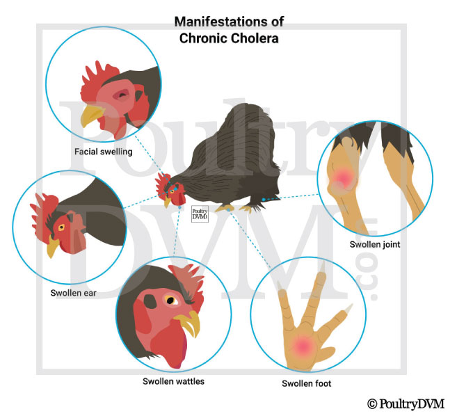
  • Chronic Form: Presents as a localized infection (swelling, inflammation, and abscess) of the wattles, sinuses, foot pad, sternal bursa, joints (leg or wing), or ears. If the ears are involved, infected birds may exhibit wry neck (torticollis) from involvement of the middle ear. Sometimes tracheal rales and dyspnea may occur secondary to respiratory tract infections. This form of FC may last 3 to 4 weeks and may sometimes persist for years, even after receiving treatment.
Manifestations of Chronic Fowl cholera in Chickens

Transmission


FC is spread via horizontal transmission through direct or indirect contact with infected birds. It is introduced to flocks through wild birds, rodents, predator attacks (especially involving domestic dogs or cats), newly introduced carrier birds, or fomites (contaminated equipment, etc). P. multocida can enter through mucous membranes, including oral, nasal, and conjunctival, as well as through cutaneous wounds. Detailed routes in which FC is spread include:
  • Direct contact with infected birds: Secretions made from infected birds, requiring close contact with one another.
  • Ingestion: Most common route, involving contamination of the environment, feed, or water with feces from infected hosts.
  • Predator attacks: Non-fatal predator attacks from wild or domestic animals (dogs, cats, and raccoons are known to be carriers of high amounts of the bacterium in their oral cavities and underneath their nails). Any chicken that has been in a predator's mouth or scratched by a predator should be treated immediately with appropriate antibiotics.
  • Fomites: Contamination of equipment, clothing, cages, feeders, etc.
  • Aerosol form
P. multocida can persist in the environment for weeks after an outbreak. Chronically infected carriers play a major role in the spread of this disease, and infected birds can remain carriers for life.

Clinical Signs

Lethargy
Fever
Reduced appetite
Ruffled feathers
Yellowish-gray diarrhea
Mucous discharge from the mouth
Increased respiratory rate
Swollen wattles, sinuses, joints, sternal bursae, ears, or footpads
Lameness
Darkened, purple head and comb (cyanosis)
Wry neck (torticollis)

Diagnosis

  • History
  • Clinical signs
  • Lab tests

Reported Cases

  • Case 1: Cellulitis of the head and conjunctivitis  in a Multiple flocks Fowl cholera due to Pasteurella multocida caused the deaths of chickens and wild waterfowl in multiple locations. P. multocida was the cause of cellulitis of the head and conjunctivitis in a backyard chicken, and pneumonia and airsacculitis in 40- to 60-week-old layer hens. Ref

  • Case 2: Fowl Cholera in a Turkey A 9-mo-old male Bronze turkey died on April 7, 2018, and was presented to the California Animal Health and Food Safety Laboratory System (CAHFS)–Turlock branch, on April 10 for postmortem examination. The turkey had been rescued 1 month previously, and resided at an animal sanctuary with one other adult male turkey. The 2 turkeys were housed together in an outdoor paddock with access to shelter and perches. They were separated into neighboring paddocks after aggressive behavior caused injury to the submitted turkey’s head, snood, and feet; however, fighting continued through the fence. Two days before death, the turkey was depressed and anorexic, and received 15 mg of meloxicam orally as recommended by the veterinarian. The submitter reported continued depression and anorexia, with acute collapse and death. Ref

  • Case 3: Fowl cholera in a Turkeys Fowl cholera due to Pasteurella multocida was diagnosed in 13-week-old Tom turkeys from a flock of approximately 5,000 birds. The flock experienced an acute onset of lethargy, lateral recumbency and increased mortality. Postmortem examination of a few turkeys revealed classical lesions of severely consolidated lungs with fibrin on the pleura but also enlarged and dark livers with pale foci of necrosis scattered throughout. P. multocida was isolated from lungs and livers Ref

  • Case 4: Fowl Cholera in a Turkeys Fowl cholera caused respiratory signs and increased mortality in a house of 10-week-old turkeys. Necropsy revealed air sacculitis, pneumonia, splenitis and arthritis. Pasteurella multocida, the agent of fowl cholera, was isolated from multiple tissues of these birds. Ref

  • Case 5: Avian cholera in a Turkeys Avian cholera (>em>Pasteurella multocida) was diagnosed as the cause of high flock mortality in which 40 turkeys in a flock of 60 died over a 10-day period. All turkeys developed black/dark red lesions on the head skin, diarrhea and lethargy prior to death. Two turkeys were submitted. Caseous cellulitis was associated with the head skin lesions which were characterized as infarcts. P. multocida was isolated from liver, lungs and subcutaneous tissues. Ref

  • Case 6: Severe infraorbital sinusitis in a Pheasants Pasteurella multocida, somatic serotype 6, was isolated from the infraorbital sinuses of 8-wk-old ring-necked pheasants with severe sinusitis. In addition, Escherichia coli, Pasteurella haemolytica-like bacteria, Mycoplasma gallinaceum, and Mycoplasma glycophilum were also isolated from some of the sinuses. Clinical signs appeared 3 days after placement on the grow-out ranch. The sinusitis consisted of severe unilateral or bilateral distention of the sinuses by mucoid to caseous exudate. Mortality and morbidity were low. Birds responded to treatment with tetracycline after proper medication procedures. The source of the infection was not determined, though possible sources include the brood ranch, wild animals, or wild waterfowl. Ref

Treatment

NameSummary
Supportive careIsolate the bird from the flock and place in a safe, comfortable, warm location (your own chicken "intensive care unit") with easy access to water and food. Limit stress. Call your veterinarian.
ProbioticsProviding chickens multi-strain probiotics may help improve intestinal health while attenuating inflammatory reaction, clinical signs and mortality.R Reuben et al., 2021
Oxytetracycline5 mg/kg SC, IM q12-24h or 2500 mg/L drinking water and 2500 mg/kg feed. Must be given simultaneously in both feed and water to be effective. Do not use outdated medication, because it becomes nephrotoxic. Do not give at the same time as giving products or food containing Al, Ca, Mg, or Fe as it will reduce or alter absorption of the drug.B Speers
Norfloxacin8-10 mg/kg PO q24h or 100 mg/L drinking water for 5 days. Not approved by the FDA for use in food-producing birds.B Speers
Tylosin15-30 mg/kg IM q6-12hB Speers
Sulfamethazine0.5 to 1% in food or 0.1% in water for at least 5 days. Infection may recur.B Speers
Streptomycin20-50 mg/kg IM q12-24h. May be nephrotoxic or neurotoxic.B Speers
Penicillin50,000 IU/kg IMB Speers

Support

Prevention

  • Attenuated live vaccines administered in drinking water
  • Prevent rodents, wild birds, and other animals from accessing or dropping their feces within coops and outdoor enclosures
  • Biosecurity

Scientific References

Age Range

Fowl cholera occurs in chickens of all ages.

Risk Factors

  • Wild bird feeders
  • Rodent populations
  • Survivor of a recent predator attack (especially from cats or dogs)
  • Introduction of a new bird to the flock
  • Stressors, such as changes in environment, nutrition, and injury, can affect the course of the disease.

Seasonality

WinterSpringSummerAutumn