Veterinary advice should be sought from your local veterinarian before applying any treatment or vaccine. Not sure who to use? Look up veterinarians who specialize in poultry using our directory listing. Find me a Vet
Thymoma

A thymoma is a localized tumor which originates from the epithelial cells of the thymus, a lymphoid organ. They can be benign or malignant. Site of metastases may occur in the lungs.
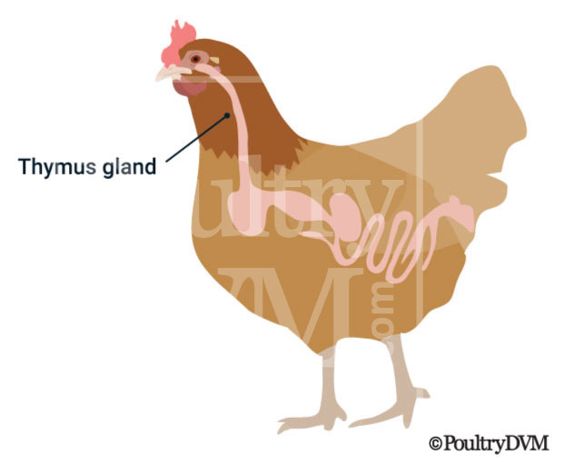
Thymus gland location in chickens
Because thymomas involve the thymic tissue, they may be found anywhere in the subcutis of the chicken's neck, between the thoracic inlet and the mandible. Tumors can appear cystic and hemorrhagic. They may present as an enlarging mass.

There have been several cases of thymoma reported in chickens. Depending on the location of the mass and health status of the chicken, surgical removal may be possible.

Clinical Signs

Subcutaneous mass on neck
Depression
Weight loss
Difficulty swallowing

Diagnosis

  • Physical exam
  • Blood tests
  • Cytology
  • Biopsy
  • CT Scan

Reported Cases

  • Case 1: Thymoma in a Chicken. A poorly differentiated squamous cell thymoma was found in a chicken in association with a lymphoma caused by Marek's disease. Immunohistochemical techniques with different tumour markers were applied in order to determine the histogenesis of the thymic tumour cells. A positive immunore action of the tumour cells to cytokeratin, considered a specific marker for epithelial tumours, confirmed the diagnosis of a thymoma. Ref
    Primary tumor site: thymusSites of Metastases: none

  • Case 2: Thymoma in a Macaw. A 49-year-old female scarlet macaw was presented with a mass in the cervical region. The bird showed no evidence of pain or discomfort, it swallowed food normally, and its breathing pattern was unremarkable. Results of contrast radiographic imaging revealed a well-demarcated mass in the right cervical region not related to the crop. After surgical resection and histopathologic evaluation, a malignant nodular stage of an epithelioid cortical-type thymoma was diagnosed. One year later, metastasis of the thymoma to the lung was diagnosed and confirmed at postmortem examination. Ref
    Primary tumor site: neckSites of Metastases: lung

  • Case 3: Thymoma in a Owl. A 12-year-old male burrowing owl was presented for evaluation of a mass in the right cervical region. A thymoma was diagnosed after surgical resection and histopathologic evaluation. Extensive adherence of the thymoma to the esophagus and suspected invasion into the right jugular vein contributed to a poor postsurgical outcome. Ref
    Primary tumor site: neckSites of Metastases: none

  • Case 4: Thymoma in a Robin. A mixed thymoma was diagnosed in a 15-year-old female American robin that exhibited poor body condition, dysphagia, hyporexia, and depression. A 1.5-cm subcutaneous nodule was present in the cranio-ventral cervical region, which had been noticed by the owner 15 days before presentation. On cytologic evaluation of a fine-needle aspirate, well-differentiated lymphocytes were observed. Surgical excision was elected; however, the mass was firmly attached to the esophagus and the jugular vein, and the attempt at excision resulted in fatal hemorrhage. On histologic examination of the mass, small, well-differentiated lymphocytes were observed mixed with neoplastic reticular cells and Hassall's corpuscles. On immunohistochemical analysis, the cytoplasm of 80% of the reticular cells showed abundant detectable brown antigen binding with pancytokeratin staining, and most lymphoid cells showed detectable antigen in the cytoplasm by using CD3 antibodies. The cytologic, histopathologic, and immunohistochemical features of the neoplasm in this robin were consistent with a mixed thymoma. Ref
    Primary tumor site: neckSites of Metastases: none

  • Case 5: Thymoma in a Sparrow. Thymoma is described in a 3-year-old female Java sparrow. Histopathologically, the subcutaneous mass from the cervical region consisted of epithelial cells characterized by large polyhedral cells and spindle cells, which sometimes showed squamous differentiation, and small lymphocytes without apparent atypical forms. Immunohistochemically, the epithelial cells possessed cytokeratin in their cytoplasm and proliferating cell nuclear antigen. Ref
    Primary tumor site: neckSites of Metastases: none

  • Case 6: Thymoma in a Chicken. A rooster of the Barred Rock breed, aged two to three years and in good body condition, was selected to be killed. His neck was cut across rather close to the thorax, and the cutting instrument severed also the posterior portion of a large tumorous mass which occupied the ventral aspect of the cervical region extending from the thoracic aperture forward for a distance of 10 cm. The abundant plumage had prevented this large tumor from being seen until the neck had been severed from the body. The mass, which weighed approximately 250 gm., had encroached on the wall of the crop and pushed this organ to one side. The dimensions of the tumor were 10 by 5 by 4 cm. It was firm, homogeneous and of a dirty flesh color, with occasional irregular foci of necrosis. The mass was situated subcutaneously and was freely movable in the connective tissue elements ventral to the trachea and the cervical vertebrae. Careful macroscopic examination failed to reveal metastatic tumors in any of the organs or tissues. Ref
    Primary tumor site: neckSites of Metastases: none

Treatment

NameSummary
Supportive careIsolate the bird from the flock and place in a safe, comfortable, warm location (your own chicken "intensive care unit") with easy access to water and food. Limit stress. Call your veterinarian.
Surgical removal of massMay be possible depending on the location.

Support

Prognosis

Poor

Scientific References

Age Range

More likely to occur in older birds.