Veterinary advice should be sought from your local veterinarian before applying any treatment or vaccine. Not sure who to use? Look up veterinarians who specialize in poultry using our directory listing. Find me a Vet
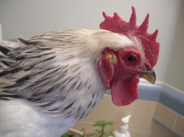
Ear Infections
Other Names: Otitis, Ear Canker
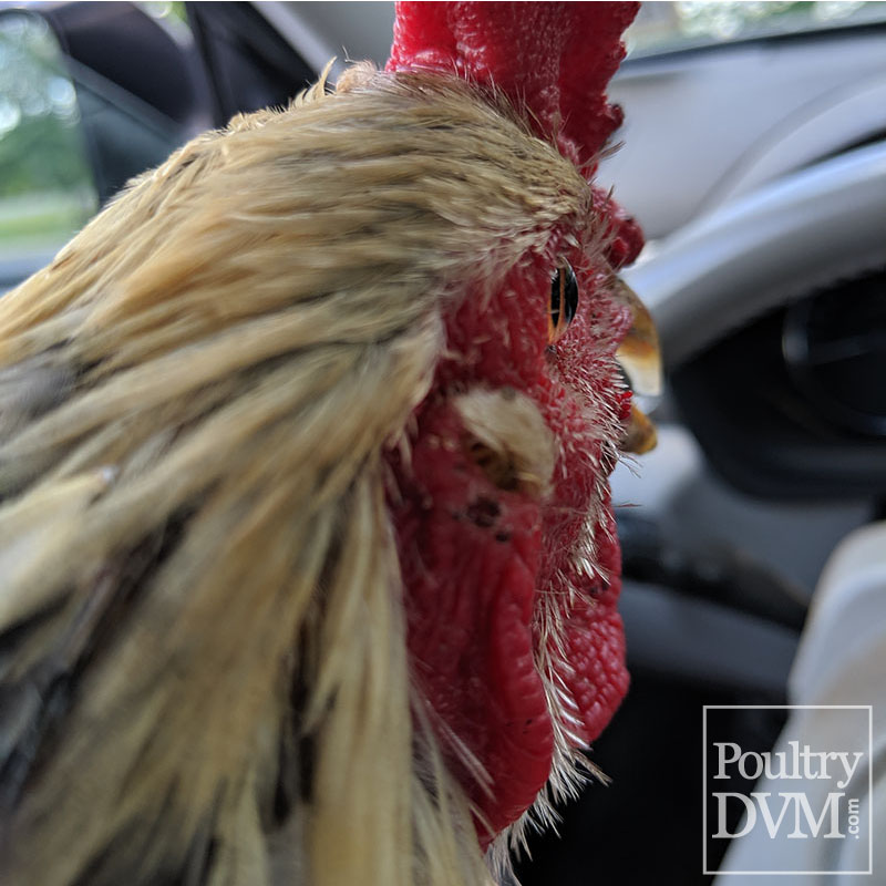
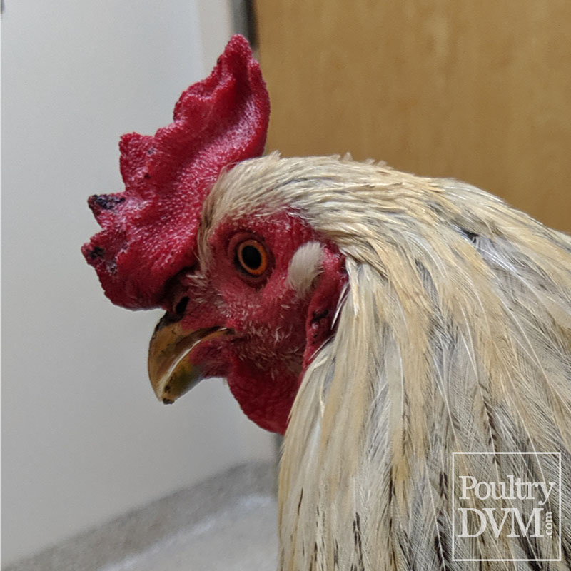
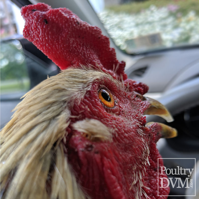
Chickens have ears which are used for hearing and balance. They are composed of an external part which is hidden by a tiny clump of stiff feathers, a middle part with an air-filled tympanic cavity, and an inner part, a complex structure with the membranous labyrinth. Ear infections can affect any or all portions of the ear.
Inner ear infections (Otitis interna): The inner ear is the sensory receptor for both sound and equilibrium. It consists of the cochlear organ and a vestibular organ. The chicken's inner ear is considered to be a part of their nervous system and helps the chicken with balance. This is why inner ear infections can cause neurological signs in affected chickens, in the form of head tilt, loss of coordination and balance, and torticollis (wry neck). Inner ear infections are most likely caused by a viral infection and are much more difficult to treat.
Middle ear infections (Otitis media): The middle ear is an air-filled tympanic cavity containing a muscle, ligaments, the tympanic membrane, the cochlear window and a rod-like bone (ossicle) known as the columella. Middle ear infections are usually caused by a chronic bacterial infection or tumor. Bacteria most commonly isolated include: Enterococcus faecalis, Escherichia coli, Pasteurella multocida, and Pseudomonas aeuroginosa. Bacteria can sometimes enter the chicken's ear through a small opening in the roof of the mouth, referred to as the infundibular cleft. The infundibular cleft opens directly into the chicken's auditory canals of the ears. These infections tend to be a little trickier to treat, as many of the invading organisms are considered to be opportunistic and highly resistant to many antibiotics. Thus, its often best to conduct antibiotic sensitivity testing prior to starting the bird on any treatment, in order to select the most effective antibiotic against the invading organism.
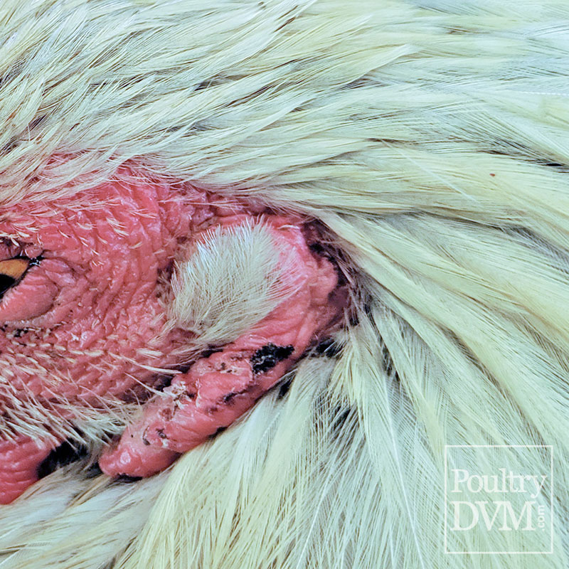
Outer ear infections (Otitis externa): Inflammation of the external ear may be caused by bacterial or fungal organisms. Pruritus may be present causing the bird to scratch their ear or rub their head often. The ear opening is often red and swollen, and the stiff clump of feathers covering the ear opening may be matted with discharge. The most frequent bacteria isolated from these infections include Pseudomonas aeuroginosa, Klebsiella spp, Enterobacter spp, and Kocuria kristinae.
Case 1: Otitis interna in a Bustard A captive juvenile little bustard was presented for acute onset of right head tilt and right circling. The bird failed to respond to supportive care and systemic antibiotic therapy. A bilateral granulomatous and fibrinoheterophilic otitis interna due to Pseudomonas aeruginosa was diagnosed postmortem by histopathologic examination and bacterial culture. Ref
Case 2: Otitis and cranial osteomyelitis in a Partridges A flock of red-legged partridges developed neurological signs, consisting of abnormal head position, torticollis (wry neck), had difficulty standing (ataxia), and difficulty walking or flying. Pathological, microbiological and molecular genetic data supported an association with Ornithobacterium rhinotracheale (ORT) infection. Clinical signs persisted for several days and were accompanied by weight loss leading to death. Morbidity was approximately 20% and most birds died if untreated. Lesions were mainly characterized by a severe osteomyelitis of the cranial bones and purulent inflammation of the external, middle and inner ears. O. rhinotracheale was isolated from ear samples, skull and brain stem in pure culture. Genetic characterization by pulsed-field gel electrophoresis of the clinical isolates showed that the outbreak was caused by a single strain of ORT. Ref
Case 3: Otitis and meningoencephalitis in a Chickens In March 2017, the Turlock branch of the California Animal Health and Food Safety laboratory system encountered an unusual clinical and pathologic presentation of infectious coryza in 6 live, 29-d-old, commercial broiler chickens that were submitted for diagnostic investigation. Antemortem evaluation revealed severe neurologic signs, including disorientation, torticollis, and opisthotonos. Swollen head–like syndrome and sinusitis were also present. Histologically, severe sinusitis, cranial osteomyelitis, otitis media and interna, and meningoencephalitis were noted, explaining the clinical signs described. A. paragallinarum was readily isolated from the upper and lower respiratory tract, brain, and cranial bones. Infectious bronchitis virus (IBV) was also detected by PCR, and IBV was isolated in embryonated chicken eggs. Based on sequencing analysis, the IBV appeared 99% homologous to strain CA1737. A synergistic effect between A. paragallinarum and IBV, resulting in exacerbation of clinical signs and increased mortality, may have occurred in this case. A. paragallinarum should be considered among the possible causes of neurologic signs in chickens. Ref
Case 4: Otitis Externa in a Lovebird A 5-year-old lovebird was presented with scaly crusts around both external ear openings and exudate present around the left ear. The bird had been treated with ivermectin and enrofloxacin without success. A pure culture of Corynebacterium kroppenstedtii was isolated from both ears. After susceptibility testing, a treatment of an acetic and boric acid solution administered topically 3 times daily was prescribed. The scaly appearance disappeared after 14 days of treatment and C. kroppenstedtii could not be re-isolated. Ref
Case 5: Otitis interna in a Turkeys Otitis interna was diagnosed in five 9-to-21-day-old turkey poults with clinical signs of paralysis, opisthotonus, torticollis, blindness, and increased mortality. Gross and microscopic lesions in the poults included omphalitis, typhlitis, hepatitis, meningoencephalitis, ophthalmitis, neuritis and ganglionitis of the vestibulocochlear nerve, and otitis interna. Salmonella enterica arizonae was isolated from the brains, eyes, intestines, yolk sacs, and livers of poults. Birds with otitis interna also had meningoencephalitis. It is most likely that the S. enterica arizonae infection spread from the brain to the internal ears through the vestibulocochlear nerve. Ref
Case 6: Otitis media in a Goose A 20-year-old Chinese goose presented for severe left-sided head tilt and circling to the left. Peripheral vestibular disease associated with otitis media extending into the left quadrate bone was diagnosed by magnetic resonance imaging and computed tomography. Otoscopy confirmed a ruptured tympanic membrane, and a brainstem auditory evoked response test confirmed loss of hearing in the affected ear. Surgery to remove the caseous material and long-term medical therapy improved the bird's head tilt and quality of life Ref
Case 7: Otitis media in a Falcon A 7-week-old male Saker falcon died with a history of severe refractory dyspnoea and respiratory signs. Microscopical lesions included moderate to severe lymphoplasmacytic inflammation of the middle ears, conjunctivae, third eyelids, choanae, salivary glands of the tongue, turbinates, larynx, trachea, syrinx and bronchi. The lesions were associated with variable numbers of Cryptosporidium spp., further confirmed by transmission electron microscopy and in-situ hybridization. Cryptosporidium baileyi was identified by DNA sequence analysis. Ref
Case 8: Pox and Otitis externa in a Backyard chickens Avian poxvirus caused severe otitis externa and media in a backyard flock hen with possible secondary bacterial infection. Ref
Isolate the bird from the flock and place in a safe, comfortable, warm location (your own chicken "intensive care unit") with easy access to water and food. Limit stress. Call your veterinarian.
Treatment depends on the cause, location and severity of the infection.